

CT scans offer a glimpse into lives of 3 Egyptian mummies
Radiologists’ expertise contributes to new exhibit at Saint Louis Art Museum
 Matt Miller
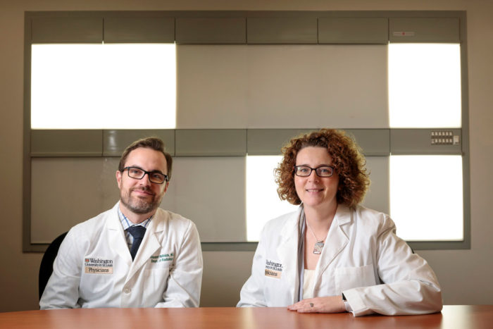
Vincent Mellnick, MD, (left) and Michelle Miller-Thomas, MD, radiologists at Washington University School of Medicine in St. Louis, conducted computerized tomography (CT) scans of three Egyptian mummies. They recently discussed their findings at the Saint Louis Art Museum. The museum has a new exhibit featuring the mummies.
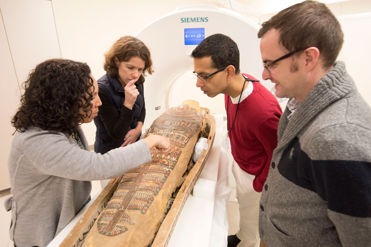
Radiologists at Washington University School of Medicine in St. Louis recently celebrated a housewarming of sorts for three former patients, each of whom is thousands of years old.
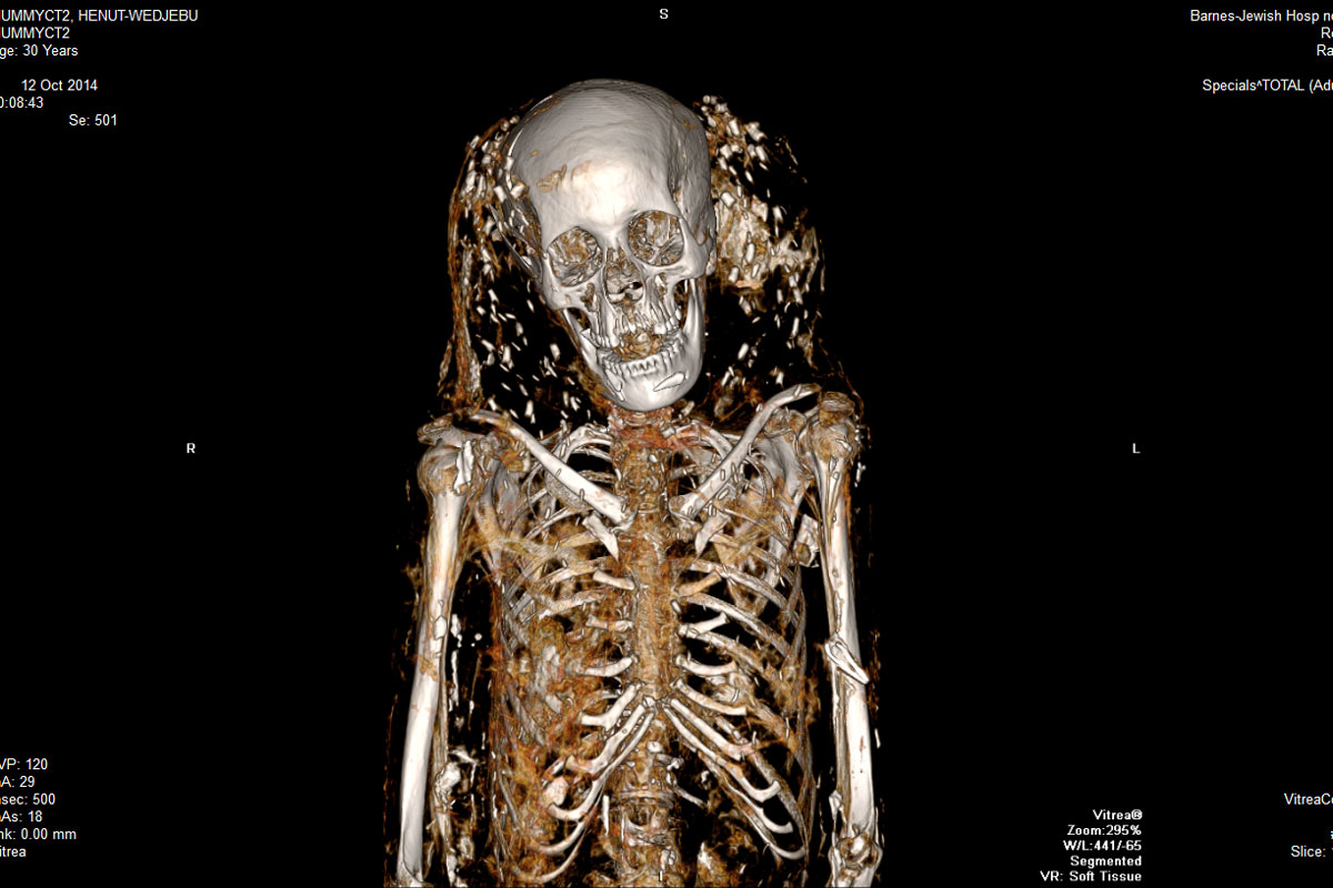
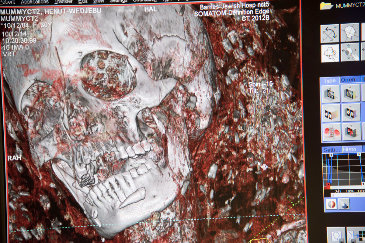
The mummified remains of Henut-wedjebu, Padi-menekh and Amen-nestawy-nakht have settled into their new, permanent exhibit space in the Saint Louis Art Museum’s Ancient Egyptian Art Gallery. The third-floor exhibit opened in December and includes hieroglyphics, artifacts and a 55-inch interactive touchscreen for visitors to explore computerized tomography (CT) scans of one of the mummies, Amen-nestawy-nakht. The images were taken in 2014 by radiologists at the university’s Mallinckrodt Institute of Radiology (MIR).
To mark the occasion, Vincent Mellnick, MD, an associate professor of radiology and chief of the abdominal imaging section, and Michelle Miller-Thomas, MD, an assistant professor of radiology, gave a talk Feb. 16 at the art museum, in which they discussed the findings of the scans with Lisa Çakmak, PhD, the museum’s associate curator for ancient art. In 2014, Sanjeev Bhalla, MD, a professor of radiology and chief of the cardiothoracic imaging section, led the university’s team of radiologists in the mummy scanning project.
 Matt Miller
The radiologists said they will continue to work with the art museum and other experts worldwide who specialize in Egyptology, postmortem decay and other areas that might shed light on the mummies’ ancient cultures.
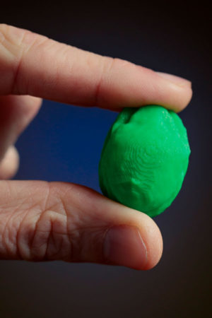
Using data from the scans and a 3-D printer, the radiologists made a copy of a scarab amulet discovered in the chest of Amen-nestawy-nakht, a mummy owned by the museum. The other two mummies are on loan from Washington University’s Mildred Lane Kemper Art Museum.
The model of the mummy’s amulet is etched in brightly colored plastic. It resembles a dung beetle belonging to the species Scarabaeus sacer, and it is similar in size to such a beetle.
The radiologists answered questions about their findings:
Can you tell us more about the scarab amulet?
Miller-Thomas: Amulets in the shape of scarab beetles were very popular in ancient Egypt. The beetle symbolized a rising and rebirth, and the scarab amulet was commonly placed over the heart and covered within the wrappings. The scans revealed the scarab amulet near Amen-nestawy-nakht’s heart.
What else did you find interesting?
Miller-Thomas: I’m a neuroradiologist so I examined the brains and skulls. Most lay people believe the brain is removed during the mummification process. It was for Amen-nestawy-nakht, a male from around 900 B.C. His brain cavity was filled with linen. Padi-menekh’s brain also was removed, and his skull was filled with a liquid resin, which has hardened. He is from the third century B.C. But Henut-wedjebu, a female from around 1300 B.C., still had her brain. It was fascinating. The mummies’ lives span over thousands of years, and the scans showed that the process of mummification had changed over the years.
We also learned about social status. Henut-wedjebu was of the highest status. We know this in part because her coffin was ornate, which indicated more wealth. Compared with the other two mummies, the CT scans showed that her teeth were in better shape and she had less dental disease. This may be a reflection of her higher status in that she had better access to food and oral care.
 Washington University
Why were CT scans ideal in learning more about the mummies?
Mellnick: The general public tends to view mummies solely as pieces of art. But, in fact, mummies are human remains. Modern imaging equipment allows us to learn more about these extinct societies without harming the bodies. It allows us to be respectful of their culture by preserving intended rituals surrounding the afterlife. Physically opening the sarcophagi was not an option to be considered.
Did it feel like the mummies were actual patients?
Bhalla: For me, the mummies were like patients. Gil Jost (a professor emeritus and former head of MIR) organized the project with the art museum. He stressed dignity and respect when we created the mummy-imaging team. He wanted us to approach them with the same thoughtful approach we do our living patients. That philosophy guided us into subspecialist review and a careful assessment with the generation of reports.
 Washington University
How will the radiology team continue to learn more about the mummies?
Miller-Thomas: We are continuing to work with the art museum and using images to consult with various experts who may offer additional insights. For instance, the scans indicated a fracture in Henut-wedjebu’s skull. I consulted with a coroner from the St. Louis County Office of the Medical Examiner. She said the pattern of the injury indicated that it happened after death, maybe because of the way her body was handled during mummification. Her joints also were dislocated, perhaps in the straightening of the body. Amen-nestawy-nakht had disruption of the spine and thorax. It most certainly occurred postmortem, possibly caused by rough handling or storing the body upright.
How has the mummy-imaging team benefited the radiology department?
Miller-Thomas: The opportunity to collaborate closely with co-workers has created friendships and strong working relationships across the department’s subspecialties.
Mellnick: It would be easy for Michelle and I, or Sanjeev and I, to go weeks, if not months, without working together. But the familiarity we have developed makes it easy to collaborate, and that helps clinical practice.
Bhalla: The most rewarding aspect was the teamwork that went into the project. Every person had value and diversity of thought and education. The result is a stronger overall team.
 Washington University

