

Mummies visit the Medical Center
Photos: Ancient Egyptian mummies arrive on campus for state-of-the-art CT scans
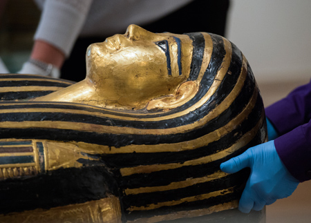
Henut-Wedjebu, the “Mistress of the House and Songstress of Amun,” was an upper-class woman who lived in the 14th century B.C. The mummies were prepared for transport at the Saint Louis Art Museum by a team of expert art handlers and museum staff. <em>Photo by Joe Angeles.</em>
Washington University radiologists welcomed a trio of special guests to the Center for Advanced Medicine this month when three Egyptian mummies arrived for computerized tomography (CT) scans, also called CAT scans. Follow their journey below, or read more in the Record.
The mummy of Henut-Wedjebu was one of three taken to Washington University Medical Center to be examined in a CT scanner.
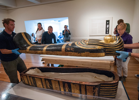
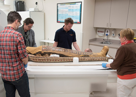
Henut-Wedjebu’s mummy and that of Pet-Menekh belong to Washington University’s Mildred Lane Kemper Art Museum but are on long-term loan to the Saint Louis Art Museum. The third mummy scanned – that of Amen-Nestawy-Nakht, a male priest believed to have lived in the 9th or 10th century B.C. – is owned by the Saint Louis Art Museum. Lifting the lid of Henut-Wedjebu’s sarcophagus is John Woodworth of US Art. Jan Hessel of Kemper Art Museum is on the right. Photo: Joe Angeles
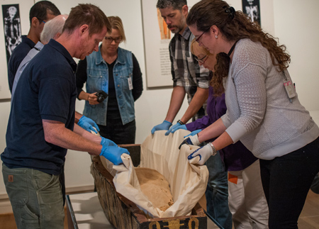
The mummies were prepared for transport at the Saint Louis Art Museum by a team of expert art handlers and museum staff. Clockwise from left are John Woodworth, art handler, US Art; Paul Haner, paintings conservator, Saint Louis Art Museum; Emilio Maldonado, art handler, US Art; Alice Boccia Paterakis, objects conservator; Ron Weaver, exhibition preparator, Kemper Art Museum; Jan Hessel, facilities manager and preparator, Kemper Art Museum; and Diane Mallow, assistant registrar, Saint Louis Art Museum. Photo: Joe Angeles
The Kemper Art Museum mummies were first X-rayed at the medical center in the 1960s. However, imaging technology has improved significantly, leading researchers to seek more detailed scans.
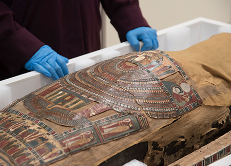
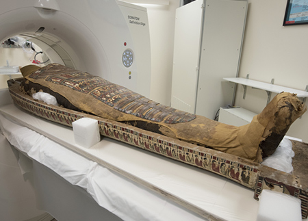
Pet-Menekh is thought to have been a priest who lived in the 4th or 3rd century B.C. His coffin is decorated with hundreds of hieroglyphics as well as images of the goddesses Isis and Nut. Photo: Robert Boston
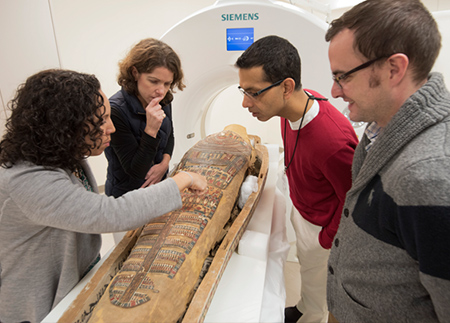
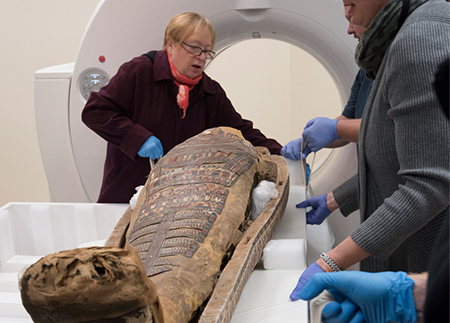
Examining Pet-Menekh’s mummy, from left, are curators Lisa Çakmak, PhD, of Saint Louis Art Museum, Karen Butler, PhD, of Kemper Art Museum, and School of Medicine radiologists Sanjeev Bhalla, MD, and Vincent Mellnick, MD. Photo: Robert Boston
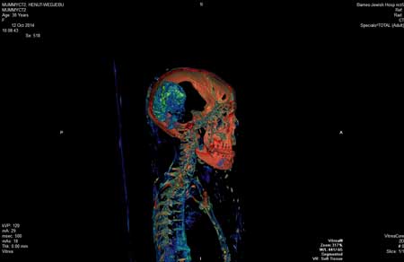
Today’s CT scans allow researchers detailed examination of the mummies in 3-D, without harming their remains.
CT scanners use special equipment that emits a narrow X-ray beam to obtain images from different angles around the body, generating 3-D images that can show the skeleton, tissues and organs. Photo of scan: Robert Boston
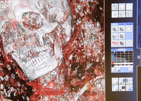
The computer adds color to the 3-D images to exaggerate differences in tissue. Photo of scan: Robert Boston
School of Medicine radiologists and museum staff hope the scans will teach them more about the mummies and the societies in which they lived.
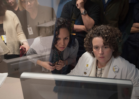
Michelle Miller-Thomas, MD, assistant professor of radiology (seated), studies the CT scans as Lisa Çakmak, PhD, of Saint Louis Art Museum leans in for a closer look. Photo: Robert Boston
Each mummy’s sarcophagus was guided safely into the scanner, thanks to careful work by the art handlers and medical team.
Positioning Pet-Menekh’s mummy from the left are Ron Weaver, exhibition preparator at Kemper Art Museum; Diane Mallow, assistant registrar at Saint Louis Art Museum; John Woodruff, an art handler with US Art; and Jan Hessel, facilities manager and preparator for the Kemper Art Museum. Photo: Robert Boston
Jan Hessel (left) of Kemper Art Museum helps position Pet-Menekh’s mummy for a CT scan. Photo: Robert Boston
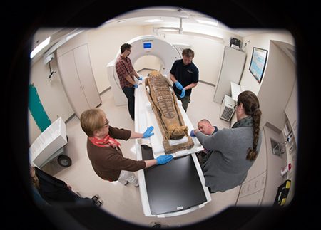
Staff from Kemper Art Museum, the Saint Louis Art Museum and US Art position Pet-Menekh’s mummy before it is examined. Photo: Robert Boston
More than 2,000 years after his body was wrapped in bandages, the mummy of Pet-Menekh is eased into a state-of-the-art CT scanner at Washington University Medical Center. Photo: Robert Boston
Researchers expect to share detailed results of the scans in December.
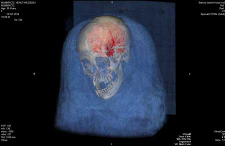
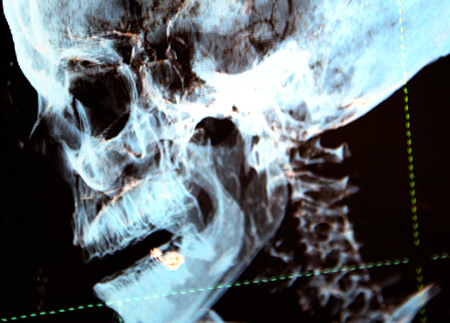
It was already known that Henut-Wedjbu was buried with her brain intact. Early findings of the scans show she still has her heart and lungs as well. In many mummies these organs were removed prior to burial. Image: Washington University

Radiologists also discovered that Henut-Wedjebu’s mummy has a collection of objects around her head. It appears to be a headdress or embellished shroud, but it could be packing material or debris. Image: Washington University
Images of the CT scans themselves may be incorporated into a reinstallation of the Egyptian mummies opening at Saint Louis Art Museum in 2016. Image: Washington University


