

Collaboration with SLU leads to purchase of $5 million microscope
Will allow shared access to imaging center, enhance biomedical research
 Matt Miller
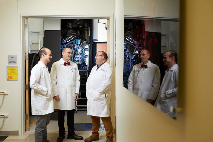
Shown at the Washington University Center for Cellular Imaging, James Fitzpatrick, PhD, (right) the center's director, visits with Enrico Di Cera, PhD, (center) chair of the Department of Biochemistry and Molecular Biology at Saint Louis University (SLU), and cryo-electron microscopy specialist Michael Rau. Washington University and SLU have signed an agreement that will extend access to the center to SLU researchers and enable the center to purchase a second cryo-electron microscope.
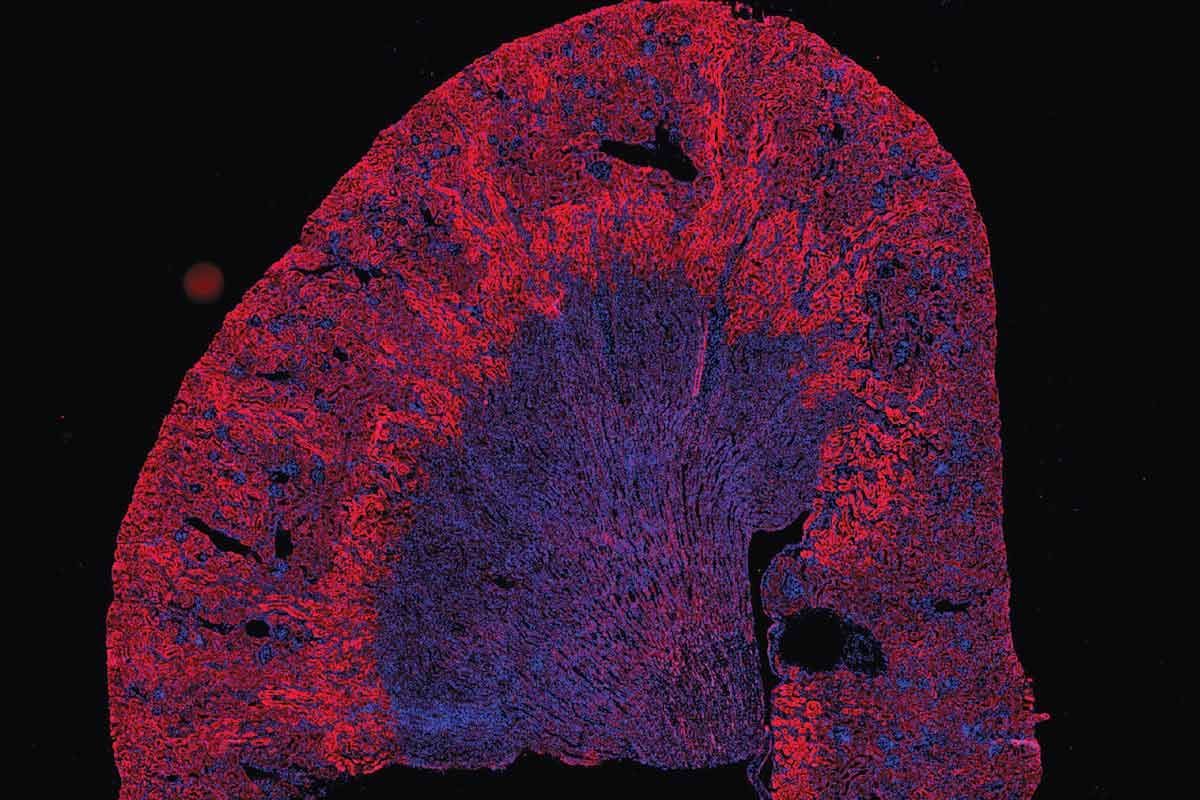
From snapshots of the molecular structure of Zika virus to 3D maps of tiny blood vessels in the lungs, images captured by the Washington University Center for Cellular Imaging (WUCCI) have provided insight into countless biological phenomena and aided investigations into myriad aspects of human health and disease.
Now, the center is poised to expand its collection of microscopes and – through an agreement between Washington University in St. Louis and Saint Louis University (SLU) – access to the center and its imaging services will be extended to SLU researchers. As part of the agreement, Washington University will purchase a new cryo-electron microscope that will be available to all users of the center.
“I am excited to begin this collaboration with SLU,” said Jennifer K. Lodge, PhD, Washington University’s vice chancellor for research. “Not only will Washington University and SLU researchers have expanded access to cutting-edge imaging technology to advance their research, I also hope that the increased opportunities for interaction between scientists at the two institutions will catalyze new ideas, new collaborations and new discoveries.”
SLU is contributing $2.5 million to the center. The contribution enables the purchase of a new $5 million cryo-electron microscope, which will be housed on the Washington University Medical Campus and maintained and supported by staff from the Center for Cellular Imaging. The microscope was ordered in December and is slated to be delivered this fall. Installation and calibration will take a couple of months, so the microscope is expected to be ready for use toward the end of the year.
“We deeply value our research partnership with Washington University as a key element of Saint Louis University’s ongoing growth as a preeminent research university to serve the St. Louis region,” said Ken Olliff, vice president for research at SLU. “I’m delighted that this new collaboration will allow SLU and Washington University scientists to access the most advanced imaging technology in the field.”
As of December, SLU researchers can use any of the microscopes and services at the center. They will pay the same rates and be given the same priority as Washington University researchers for any microscopes or services that are unavailable at SLU.
 Matt Miller
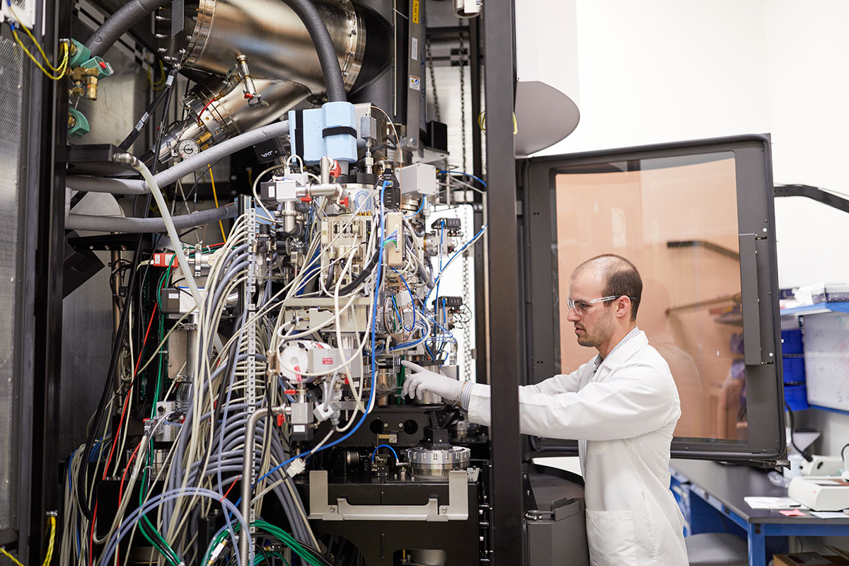
The center already hosts one of the most advanced cryo-electron microscopes available: a Titan Krios, which was installed in 2017. It has proven exceptionally popular. Images created using the microscope already have led to groundbreaking scientific discoveries.
Daved Fremont, PhD, a professor of pathology and immunology, and colleagues took a high-resolution snapshot of an arthritis-causing virus known as chikungunya, attached to the protein it uses to invade cells. The image may help researchers design drugs or vaccines to prevent or treat arthritis caused by chikungunya or related viruses.
Rui Zhang, PhD, an assistant professor of biochemistry and molecular biophysics, used the cryo-electron microscope to take the first detailed look at the inner structure of cilia, whiplike appendages on cells that are involved in a wide range of biological processes including moving mucus in the respiratory tract, propelling sperm and helping bone grow. Understanding the structure of these microscopic molecular machines potentially could answer questions about human health and disease.
“The agreement between SLU and Washington University cements a common desire to grow our footprint in structural biology,” said Enrico Di Cera, MD, the Alice A. Doisy Professor and chairman of the Department of Biochemistry and Molecular Biology at SLU.
“Acquisition of state-of-the-art technology and, most importantly, the resulting scientific interactions between SLU and Washington University faculty members will bring long-lasting benefits to our institutions. We will continue to grow our existing strengths and establish new ones with the recruitment of new talent. Last but not least, this investment from SLU comes from the Doisy Fund in Biochemistry and fits perfectly the original intent and scientific legacy of Nobel Prize winner, department founder and SLU benefactor Edward Doisy.”
Washington University and SLU each boast Doisy in their respective histories. Doisy was on the Washington University School of Medicine faculty for four years before joining the faculty at SLU.
The machine operates seven days a week, said James Fitzpatrick, PhD, director of the Center for Cellular Imaging. Fitzpatrick and Michael Rau – the center’s cryo-electron microscopy specialist – give up part of their weekends to come to the center to make sure the system is running smoothly, but the waiting list is still two months long.
The collaboration agreement gives SLU researchers access to the center for 10 years and access to both cryo-electron microscopes for 15 years. The center also boasts 19 other light, X-ray and electron microscopes and provides training and assistance with sample preparation and data analysis. Only the cryo-electron microscope has a significant wait time, so the center will be able to handle any increased demand of SLU researchers on other microscopes and services without reducing availability for Washington University researchers, Fitzpatrick said.
“Further expanding our cryo-EM capacity by the purchase of an additional cryo-electron microscope is a win-win for both SLU and Washington University,” said Fitzpatrick, who is also a professor of neuroscience and of cell biology and physiology. “It will allow us not only to decrease wait times for scientists at both institutions, but also enable us to offer new services not currently possible at the center.”
The new cryo-electron microscope will be a Glacios, a different model than the Krios already in use at the center. While the new microscope will have some of the same capabilities as the first one, it also will be able to image samples using an approach called micro-electron diffraction. The recently developed technique is valuable for visualizing molecular structures that are too small for conventional cryo-electron microscopy methods. Between the two microscopes, researchers will be able to make detailed images at nanoscale resolution of everything from single small molecules to viruses thousands of times bigger.
 Related:

