Washington University invests $25 million in imaging sciences
Initiative will feature engineering, medicine collaborations

Building on its extensive history in imaging — from individual cells and nerves to cancerous tumors and Alzheimer’s plaques — Washington University in St. Louis is launching a bold, $25 million initiative over the next five years to support its researchers as they develop innovative technologies aimed at improving science and medicine worldwide.
Initially, the Imaging Sciences Initiative — a partnership between the School of Engineering & Applied Science and the School of Medicine — will bring in more than a dozen new faculty with strengths in various aspects of imaging sciences. Both schools have their own long-standing strengths in the field, with more than 100 imaging scientists between them.
The new initiative strengthens the connection between the schools and encourages the development of new imaging technologies to diagnose and treat disease as well as study intricate biological structures, metabolism and physiology, and critical molecular and cellular processes.
“This initiative allows us to attack challenges in imaging that can only be addressed by collaborations between medicine and engineering, including developing fundamental new technologies and advanced computational methods,” said Aaron F. Bobick, dean of the School of Engineering & Applied Science and the James M. McKelvey Professor. “Washington University will further establish its place at the forefront of groundbreaking biomedical engineering and imaging research that can have an immediate impact in the world.”
Six departments in the two schools will join the initiative: the Departments of Biomedical Engineering, Computer Science & Engineering, and Electrical & Systems Engineering, at the engineering school; and the Departments of Radiology, Radiation Oncology, and Cell Biology & Physiology, at the medical school. Additional faculty as part of the initiative are planned for subsequent years.
Beyond the faculty recruiting effort, the initiative includes plans for imaging research centers focused on fundamental science and technology, as well as translational clinical opportunities. In addition, an interdisciplinary doctoral program in imaging sciences will be established.
“I am thrilled with the potential this initiative holds for pushing the boundaries of imaging technology to better diagnose and treat cancer, Alzheimer’s and other diseases that so tragically affect society,” said David H. Perlmutter, MD, executive vice chancellor for medical affairs and dean of the School of Medicine. “Welcoming new scientists to our faculty and training the next generation of imaging scientists is exciting. I am eager to see the benefits this effort will have on mankind.”
At the outset of the initiative, eight new faculty members will be hired for the 2017-18 academic year: four in Engineering and four in Medicine, with two in the Department of Radiology and one each in the Departments of Radiation Oncology and Cell Biology & Physiology. Four more faculty are expected to be added in the 2018-19 academic year.
“There is a very strong history here in imaging, and we want to maintain and build on that tradition,” said Steven C. George, MD, PhD, the Elvera & William Stuckenberg Professor of Technology & Human Affairs and chair of the Department of Biomedical Engineering. “By bringing together the vast knowledge base between basic scientists in engineering and medicine, we have the potential to change medicine as we know it.”
Joseph P. Culver, PhD, a professor of radiology, of physics and of biomedical engineering, agreed. “Imaging sciences is fundamentally an interdisciplinary science that is at the boundary between several fields. It continues to be an extremely rich area for innovation,” Culver said. “In this initiative, we are seeking applicants who will develop new imaging methods targeted to the cutting edge of biomedical research.”
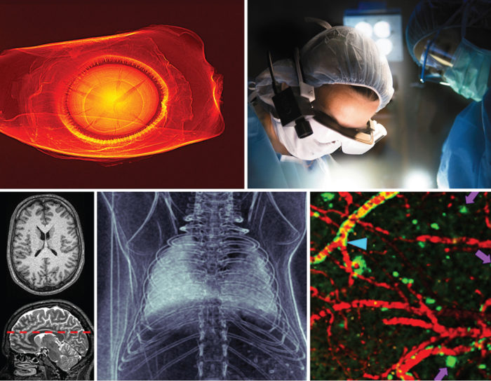
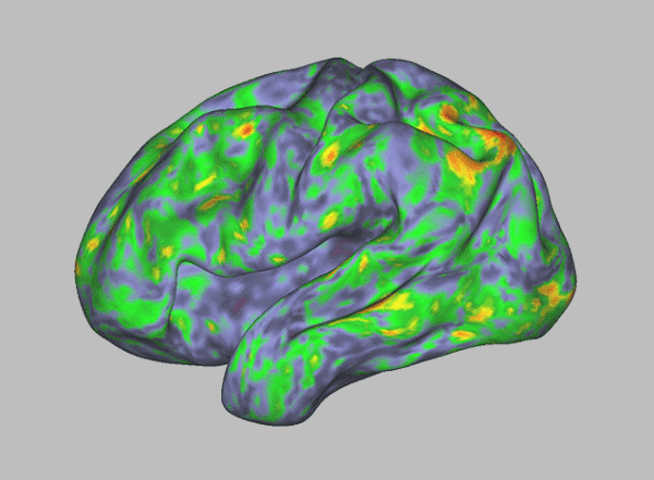
Washington University is recognized as a world leader in imaging sciences, with much of its work concentrated at the Mallinckrodt Institute of Radiology. University scientists, for example, are engaged in research to: map the myriad connections and networks in the human brain as well as regions of brain function; develop hi-tech goggles that, when used with a special dye, illuminate cancer cells; and visualize drought-related structural damage to plants that can’t be seen by the naked eye.
“We are excited to grow areas such as super-resolution microscopy, a kind of light microscopy of exceedingly tiny objects; correlative light and electron microscopy, which combine the advantages of light and electron microscopy to visualize cellular structures and processes in fine detail; and dynamic live cell imaging,” said David Piston, PhD, the Edward J. Mallinckrodt Jr. Professor and head of the Department of Cell Biology and Physiology. “To explore and develop these imaging methods, medical researchers need the help of engineers. This new initiative will allow us to grow and nurture such collaborations in ways that would not be possible by recruiting new faculty members through a single department.”
Washington University’s history in imaging dates back more than 125 years, when physicians used X-rays in a spinal surgery at the School of Medicine. The university’s first hospital was among the early leaders in adopting X-ray technology and teaching it to students. In the 1920s, Washington University researchers were the first to use X-rays to view the gallbladder. And as early as the 1930s, university researchers and physicians used imaging technologies to diagnose pulmonary and heart diseases. In the 1970s, research by Michel Ter-Pogossian, PhD, at the university’s Mallinckrodt Institute of Radiology, led to the development of the PET scanner. PET scans now are used worldwide to detect disease and as a research tool.
More recently, Washington University researchers led the development of optical probes for imaging of gene expression in cancer cells and protein-protein interactions. In 2008, Tim Holy, PhD, now professor of neuroscience, developed the Objective Coupled Planar Illumination microscope that enables rapid imaging of tens of thousands of neurons in 3-D. His work set a world record for the largest number of neurons imaged at a single time.
The pioneering work in radiation treatment planning by Jerome Cox Jr., senior professor in computer science & engineering, paved the way for systems still used today to reconstruct images from CT and PET scanners that aid in the diagnosis of cancers and cardiovascular disease. In addition, his Laboratory Instrument Computer (LINC), developed in collaboration with researchers from the Mallinckrodt Institute of Radiology, transformed biomedical research by integrating computer science with medicine.


