

New technique aids search for genetic roots of disease
Researchers can create sets of mutated DNA sequences quickly, cheaply
 Michael Worful
A new technique to cheaply and rapidly create sets of DNA fragments that include all possible genetic variants will help scientists distinguish between genetic variants linked to disease and those that are innocuous.
Some 10 million points of genetic variation are scattered across a molecule of DNA, and those variations make us who we are as individuals. But in some cases, those variants contribute to diseases, and it’s a major challenge for scientists to distinguish between harmless variants and those that are potentially hazardous to our health.
Now, researchers at Washington University School of Medicine in St. Louis have developed a new technique to cheaply and rapidly create myriad sets of DNA fragments that detail all possible genetic variants in a particular stretch of DNA. By studying such DNA fragments, scientists can more easily distinguish between genetic variants linked to disease and those that are innocuous.
The findings, published Oct. 3 in Nature Methods, allow researchers to create sets of DNA variants in a single day for a few hundred dollars. Current methods take up to a week and cost tens of thousands of dollars.
“As a pediatric neurologist who does a lot of genetic studies of kids with developmental disabilities, I frequently will scan a patient’s whole genome for genetic variants,” said Christina Gurnett, MD, PhD, the study’s senior author and an associate professor of neurology and of pediatrics. “Sometimes I’ll find a known variant that causes a particular disease, but more often than not I find genetic variants that no one’s ever seen before, and those results are very hard to interpret.”
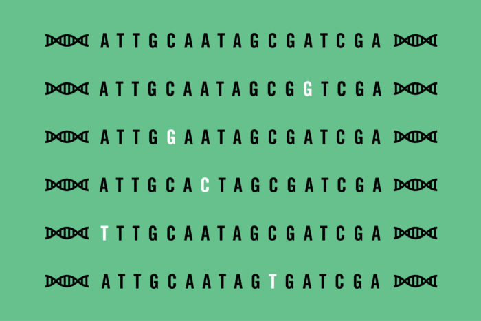
In the past, scientists tested the effect of genetic variants one by one, a laborious process. At a single point in the DNA sequence, they replaced the correct DNA letter – an A, T, C or G – with one of the other three options. Then, they translated that DNA sequence into a protein and evaluated whether the mutated protein behaved differently than the original one.
More recently, researchers have begun creating sets of hundreds of variants in which each letter in a particular DNA sequence is changed, and then testing the set all at once. Such studies have been limited, however, by the high cost of creating those sets.
Postdoctoral researcher Gabriel Haller, PhD, who was working in Gurnett’s lab, realized that he could harness common laboratory techniques and tools to create sets of DNA variants without the expensive equipment and reagents that drove up the price.
Haller copied a DNA sequence using the four standard DNA letters and a nonstandard letter known as inosine. Each copy of the sequence was identical except for one inosine, which was located at a random spot and served as a placeholder. Then, he replaced the inosine with one of the standard DNA letters, creating a single mutation in each copy of the sequence.
Gurnett and colleagues are applying this technique to genes associated with aortic aneurysms, a weakening and ballooning of the aortic wall that can be fatal. Over the long term, Gurnett envisions the creation of a catalog listing the effects of every possible variant. The speed and cheapness of the new technique make such a catalog possible.
“Then, when clinicians find a variant that’s never been seen before in one of these genes associated with aortic aneurysm, they can go through this catalog and say, ‘Yes, this mutation does have a negative effect on that protein, so it’s likely harmful’,” Gurnett said. “It would help them decide what to tell the patient. This would be one piece of the big interpretation puzzle for genetic mutations.”


