

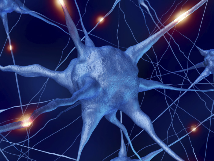
Mapping the neural networks of epilepsy
Research into the functional organization of epilepsy-altered brains may lead to early identification of best treatment option
Nearly 150,000 Americans develop epilepsy each year. For about 70 percent of patients, medication will control seizures. Surgery will prove to be the only solution for others, and a small number of patients will improve without intervention. Unfortunately, there is no crystal ball to indicate where any given patient will fall on that spectrum.
What if there was a way to know, say as early as the first or second seizure, which of these unique brains will respond to medicine and which will not?
Researchers at Washington University hope to find a way to do just that through advanced functional magnetic resonance imaging (fMRI) techniques, which can capture the complex neuronal connections between pin-head-sized patches of brain tissue.
Neurologist Luigi Maccotta, MD, PhD, used this technology to obtain finely detailed anatomical brain images and detailed maps of the activity patterns of more than 30 patients with temporal lobe epilepsy (TLE). TLE is the most common form of “focal seizures,” in which abnormal neuronal activity starts from a specific area on one side of the brain. Maccotta recruited TLE patients whose seizure onset area was already known in order to observe brain connections propagating from that point across all brain regions.
Brain activity patterns in patients with TLE were significantly different from the functional networks of normal control subjects without epilepsy—and reached far beyond the seizure onset area. Moreover, the pattern differences were so specific that it may become possible to use fMRI for early diagnosis of TLE.
Linking brain activity to optimal treatment
Now, Maccotta hopes to use the ability to assess variations in functional connections in the brain with the goal of linking specific activity patterns with best treatments. “We could dramatically improve the lives of our patients if we could determine—shortly after the onset of seizures—whether surgery is going to be their only solution, or whether medicine will work. And, if medicine is the answer, identify right away which drug is right for them.”
To do this, he plans a longitudinal study of 120 patients who have experienced only one or two seizures to date. Using the advanced fMRI techniques honed in his previous study, Maccotta will map early, seizure-generated changes in the connection patterns of individual patients. “Then, as we follow their progress and see how they respond to treatment options over time, we can look for early pattern changes that distinguish patients who will benefit from medical management from those who will not.”


