

Immune cells contribute to macular degeneration
New research shows immune cells in mice may damage or protect against age-related macular degeneration, depending on the cells’ age
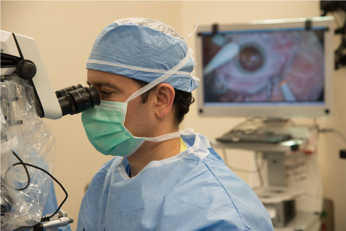
Rajendra Apte, MD, PhD, performing eye surgery.
A good deal of research suggests that inflammation can damage the retina in age-related macular degeneration (AMD). A series of studies from ophthalmology researchers at Washington University School of Medicine is revealing that immune cells called macrophages both protect the eye from damage and cause damage, depending partly on the age of those macrophages.
Macrophages are white blood cells that engulf and digest cellular debris and infectious agents, attacking foreign substances and microbes. Found in the liver, spleen and connective tissues, they also stimulate other immune cells to respond to infectious pathogens. But as the macrophage cells age, they appear to get less efficient at those jobs.
Age-related macular degeneration is the leading cause of blindness in the United States in people over 50. It accounts for more than 40 percent of blindness among the institutionalized elderly, and as baby boomers continue to age, the problem is expected to grow, with at least 8 million cases of AMD predicted by the year 2020.
There are two varieties of AMD: a “dry” form and a “wet” form. Most patients have the dry form of the disease, and although it can progress and sometimes cause severe vision loss, between 80 percent and 90 percent of the blindness and severe vision loss in AMD is related to the wet form, in which abnormal blood vessels form underneath the retina.
Leaky blood vessels
The blood vessels that characterize AMD are not like the mature vessels found in most of the body. Vessels associated with the disease don’t have normal, tight junctions but rather leak and bleed. They also tend to be located beneath the macula, the center of the retina, and when they bleed, the result is vision damage.
In several mouse studies, scientists, led by Rajendra Apte, MD, PhD, the Paul A. Cibis Distinguished Professor of Ophthalmology and Visual Sciences, have discovered that macrophages in older mice don’t block the development of those abnormal, leaky blood vessels beneath the retina as well as macrophages in younger mice.
According to Apte, whether or not the macrophages block the damaging blood vessels is related not only to the animal’s age, but to what subtype the macrophage cell happens to be.
“There are two basic types of macrophages — known as M1 and M2 — and in older mice, there are more macrophage cells with the M2 chemical signature,” he says. “The M2 cells promote abnormal blood vessel growth in the eye. In younger mice, most macrophages have the M1 signature, and those cells tend to inhibit the development of defective blood vessels.”
Apte says that the age-related shift in mice macrophages may be partly due to an increase in the levels of an immune system molecule known as interleukin-10 (IL-10). As mice get older and make more IL-10, they also end up with more M2 macrophages, elevating the risk for abnormal blood vessel formation beneath the retina.
Cholesterol, inflammation and the eye
But there’s more to it than just the type of macrophage cell. In experiments done in both mice and in human cells, Apte’s laboratory has found that as macrophage cells age, they are able to clear fewer fat deposits beneath the retina. Eventually, the macrophages become bloated with cholesterol, creating inflammation that also contributes to the formation of blood vessels, leading to the wet form of AMD.

“Ultimately, that inflammation creates a toxic mix that leads to new blood vessel growth,” Apte explains. “But the good news is that when we treat macrophages with a substance that helps clear cholesterol, we can slow the development of new vessel formation.”
Patients who have plaque build up in the arteries known asatherosclerosisoften take medications to lower cholesterol and keep their arteries clear. Apte’s research suggests some of those same drugs may be useful in patients with macular degeneration. He says the research raises the intriguing possibility that if drugs to lower cholesterol could be delivered in eye drops, it may be possible to prevent some of the vision damage caused by abnormal blood vessel growth in patients with AMD.
“We can reverse the disease cascade in mice by improving macrophage function,” Apte says. “And some of the therapies used to treat atherosclerosis target the same pathway we targeted, so it may be possible to modify drugs that already are available and to use them to deliver treatment to the eye.”
Other factors may be involved
Apte says it appears that as people age, they also make more IL-10, as well as other immune molecules that influence a drift towards macrophages that can’t stop new vessel formation. He says other factors such as smoking, uncontrolled high blood pressure, or a person’s genetic makeup also may be involved.
“The microenvironment in and around the eye seems to influence how macrophages behave,” he says. “We believe this cascade involving IL-10 and macrophages provides potential targets for therapies to prevent some of the devastating vision loss that affects so many patients with AMD and may even be useful in treating other diseases that involve abnormal blood vessel growth, such as cancer and heart disease.”


