New approach lets neurosurgeons map the brain faster, cheaper
New research at Washington University shows looking at the brain while at rest can help scientists learn about the structure and function of the brain

Mapping the brain is critical for neurosurgeons, who not only want to get a detailed fix on areas targeted for surgical repair or removal but also want to minimize risks of surgical damage to healthy brain regions that perform essential tasks.
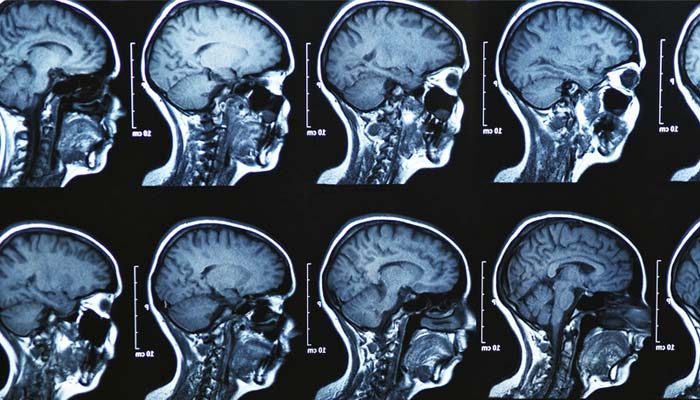
For many years, neurosurgeons have accomplished the job using a time-intensive imaging approach called functional magnetic resonance imaging (fMRI). Patients perform several simple tasks—saying their name or moving an arm, for example—while their brains are repeatedly scanned. Researchers look for areas of increased blood flow during tasks to pinpoint the brain regions involved.
Now research at Washington University shows that the same can be accomplished quicker and cheaper looking at the brain at rest.
“We had always assumed that the brain’s resting blood flow levels alone were not useful in mapping brain function,” said Eric Leuthardt, MD, associate professor of neurological surgery and director of the Center for the Integration of Neuroscience and Technology. “But then one of our colleagues, Marcus Raichle, MD, showed that looking at the resting brain is a very efficient way to learn about the functional architecture of the brain.”
Raichle learned in the late 1990s and early 2000s that blood flow to multiple brain regions rose and fell together as subjects rested in the scanner. He established that these regions were what he called “default mode networks”—collections of brain areas whose blood flow levels in the resting brain were connected for reasons scientists are still exploring. Raichle calls the technique resting-state fMRI.
Using past fMRI data describing networks in the active brain as a reference point, Raichle and colleagues were able to associate parts of the brain in resting-state networks with brain regions already linked to specific functions.
Mapping the brain at rest

Leuthardt recently has shown that resting state fMRI can be very useful for neurosurgeons to precisely map all of an individual patient’s critical brain networks—speech, motor control, and others. “It’s a super-efficient way to map the brain,” he says. “Instead of having multiple imaging sessions where patients have to do something, then rest, then repeat that over and over again, we can get all the information we need in one 15-minute session where they just lie in the scanner and rest.”
According to Leuthardt, this is particularly helpful for patients who might have trouble staying in the MRI for prolonged periods of time or hearing and following instructions during regular fMRI.
“This massively expands our ability to map patients prior to surgery, and it’s very quickly being adopted by physicians throughout the Department of Neurosurgery,” Leuthardt said.
Learn more about neurological technologies and brain function studies being developed at Washington University.


